


This disease is poorly studied, although several thousands of observations have been described with the diagnosis and subsequent treatment.
Much diversity and non -specificity of the clinical image of the varicose veins of the pelvis lead to gross diagnostic errors, which in the future affects the consequences.
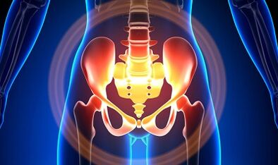
Characterization of the varicose veins of a small pelvis
The veins of the pelvis are several times longer than the arteries, which determines its great capacity.This is due to the phylogenesis of the vascular system of the pelvic region.The veins of the pelvis have high adaptation skills and is potentially predisposed to restructuring, which contributes to the formation of a densely intertwined network.
The speed and direction of the blood flow are regulated by valves, which are controlled by complex humoral mechanisms.The valves balance the pressure in different parts of the venous network.
When the valves stop performing their functions, blood stagnation develops, this leads to the pathology of blood vessels and the formation of varicose veins.The uniqueness of the veins of the small pelvis is in the fact that the wide ligaments of the uterus, which support the light of the container, can also reduce it, causing a pathology.
Causes
The pathological dilation of the veins of the pelvis may be due to the following reasons:
- Violation of blood exit paths;
- Venous barrel prison;
- Compression of collateral trunks by the changed position of the uterus, for example, in retroflexia;
- Valve ovary insufficiency (congenital or acquired);
- Obstructive postphlebetic syndrome;
- Connective tissue pathology;
- Arteriovenous angiodisplasia;
- Prolonged sitting, hard physical work;
- Varicose veins of the lower extremities;
- Pregnancy (3 or more) and childbirth (2 or more);
- Female genital area diseases (chronic salpingophorite, ovarian tumors, uterine fibroids and genital endometriosis);
- The adhesive process of the pelvic organs;
- Obesity.
Classification by degrees of the disease
In terms of expanded vein, the following degrees are distinguished:
- Up to 0.5 cm, stroke "corkscrew";
- 0.6-1 cm;
- More than 1 cm.
Options for the course of the disease
- varicose veins of the perineum and lobby of the vagina;
- Venous Pelvis Venous Blood Syndrome;
Symptoms
- The most common pain: frequent in the lower abdomen, perineum after excessive static and dynamic.The pain intensifies in the second phase of the cycle, after hypothermia, fatigue, stress, exacerbations of various diseases.
- The feeling is "not to taste", pain with sex and then.
- Dysmenorrhea - Menstruation disorders, including pain syndrome.
- Secretation, more than normal, genital tract guts.
- Blood stagnation leads to infertility, disability, pregnancy termination.
- Urine violations as a result of the expansion of the bladder veins.
Diagnosis
The diagnosis of the disease only for complaints is successful in just 10 % of cases.
The palpation of the inner walls of the pelvis makes it possible to feel oblong seals and veins nodes.When examined in the mirrors, the mucous membrane cyanosis of the vagina is visible.
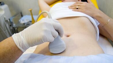
The procedure for choosing is an ultrasound study with color doppler mapping, which allows you to identify not only varicose extensions of ovarian veins, but also venous thrombosis, post -pocophble occlusion.With ultrasound, it is visible, "worms", the structure without reflection of the signal, located on the lateral surface of the uterus.
The effect of the Doppler study is based on "dye" in blue and red, venous and arterial blood, corresponding.
The ultrasonic exam device using a special program recognizes the movement of the sensor's blood and, in the other direction, calculates the speed of the blood flow and the type of glass.
But the exact definition of the vein is either the artery remains behind the doctor.The Doppler method works in almost all cases, our body dictates exceptions to the rules, since the blood that flows from the heart is not always arterial and vice versa.
Therefore, an ultrasonic diagnosis doctor sees an arterial or vessel vessel, its size, blood flow rate and many indicators that an common person does not need, but plays an important role in the diagnosis.To do this, use transvaginal and transvaginal sensors.
In 5.7% of cases, the disease is recognized by accident during the exam.Normally, the diameter of the ovarian vein is 0.4 cm.
CT and MRI have great precision.Using these methods, you can find accumulations of varicose veins in ligaments of the uterus, ovaries and around these organs.It is possible to determine concomitant pathology.
A very reliable method is a phlebographic study.
The contrast is carried out at the height of the Valsalva sample, against blood flow.This allows you to see the valve failure.
Retronezoscopy to the left, renal phlebography, super sshénica and phlegovarography on both sides are also used.These methods allow you to determine the hemodem and anatomical changes in the kidney veins and the places to fall into them.
Super Sspent phrofavarioscopy is performed through catheterization of the veins of gonads through a counteractic femur or a subclavian vein, with the subsequent introduction of contrast.
Most of the plexus blood without a varicose horn is thrown through the ovarian vein.But in conditions of hypertension, it occurs through the uterine veins not with cornillo in the internal iliac vein.The plexus of the veins, through which the output flow can occur, includes sacral and plexus of the bladder.
In the phlebo -icigography on the left sides, 3 stages of venous stagnation are distinguished in the cluster plexus of the left ovary:
- There is no exit from the left ovary plexus or is carried out along an additional short route.
- There is an additional way.
- Two additional output routes or an additional and auxiliary auxiliary are seen.
At 2 and 3 stages, the varicose veins of the right ovary cluster plexter are formed.
Laparoscopy is used for differential diagnosis.The pathologically complicated veins are in the ovary, in the direction of the round and wide ligaments.They look like large cyanotic conglomerates with a thin and tense wall.
The complexity of the diagnosis lies in the fact that the disease is often hidden behind the signs of the inflammatory process, is distinguished by clinical manifestations, masked under endometriosis, prolapse of internal organs, postoperative neuropathies and many extracturizing diseases.
Treatment
The main objective of the treatment is to eliminate reflux in the veins.In the initial stages of the disease, conservative treatment is used.In the last stages of the disease, the treatment of choice is surgery.
Conservative treatment

It consists of normalizing the tone of the veins, the improvement of hemodynamics and trophic processes.
Symptomatic treatment to eliminate individual symptoms.Non -steroidal anti -inflammatory for pain, with hemorrhage - hemostatic therapy.
The main drugs in conservative treatment are venotonic and antiplazing drugs.
Phlebotonics: improve the vascular wall tone and improve blood flow.With this disease, it is better to consult a gynecologist about certain medications.
An important method is physiotherapy exercises.
Surgical treatment
- Varicose vain veins.
- Gonad-cavalry derivation.
- Sclerosis for laparoscopy.
- Ovar western veins using endovascular x -ray methods.
Popular remedies
Since the main thing in the appearance of the disease is the weakness of the valve apparatus, all popular remedies that are used for the varicose expansion of the lower extremities veins are also used for this pathology.
The most commonly used: ordinary hazelnut, hops, orgas, horse chestnut, lion tooth root, tea fungus, sauce, oak, San Juan grass, a series, a series of flowers and many more plants.
Effective is: treatment with oak baths, brown, sauce, chamomile, pharmacy, drying herbs, San Juan grass.
Prevention
- The first thing to do if you have complaints, predictors or diseases listed above: contact a gynecologist.
- It is necessary to normalize the work regime and rest, try not to remain in a vertical position for a long time, physical tension.
- Perform exercises for "pedal" prevention, "standing battery", "feet legs"
- Adhere to the diet: Coma foods with a high content of vitamins E, R, C, try to eat only white meat, less fat, replace it with fruits, vegetables, cereals.
- Drink a sufficient amount of liquid, but not less than 1.5 liters per day.
- Shark out of excess weight, bad habits.
- Consult with the treating doctor on the use of compression linen, will improve blood outlet from the lower extremities, therefore, there will be less stagnation in the pelvis.
- Avoid bathrooms, saunas, steam rooms, hot baths.
In order not to get sick with such a difficult disease, it is necessary to follow the preventive recommendations mentioned above.Try your health as the most valuable in life.
In the slightest suspicious symptoms that cannot be undone of several days, you must consult a doctor.You must give you highly qualified help and prevent you from suffering.